


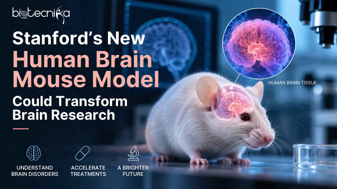
Stanford’s New Human-Brain Mouse Model Could Change Brain Research
A major scientific advancement could change the way researchers study the human brain. Scientists in the United States have developed genetically modified mice with functioning human brain cells, creating a new Human-Brain Mouse Model that could help researchers study neurological and psychiatric conditions that are difficult to reproduce in conventional animals.
According to the research paper published in Nature authored by Stanford University researchers, this advanced innovation will provide a solution to a problem that has challenged research into the workings of the brain for a while.
Eventhough, mice do not develop the same neurological or psychiatric problems that humans do. This limits our understanding of some diseases and means we cannot experiment with treatments before they enter clinical trials or effectively test them out on humans.
Prof Sergiu Pașca, the lead author of this reserach, stated that the field of psychiatry has one of the worst records of success in clinical trials. In some cases, therapies that worked well on animals proved to be ineffective in humans, indicating that existing laboratory models failed to mimic the biology of the human brain.
How was the Human-Brain Mouse Model developed?
Firstly, scientists modified the mice to have virtually no cortex of their own.
The cortex is the external part of the brain. Sometimes it is called grey matter. This is where some processes like memory or high-level thinking occur.
Then scientists used human skin cells and turned them into cells that could form brain-like tissues – organoids.
Organoids are not miniature brains. These are live tissue clusters of cells with the ability to create some features of human brain tissue development.
Next, scientists grafted the organoids into the brain of the modified mice.
Once grafted, the human cells started developing and forming contacts both with the existing brain circuit of the animal and with other parts of the brain and spinal cord.
The cortex created by the graft was not the same as a human or mouse cortex. The healthy cortex is always formed in layers, while the grafted one looked less organised.
However, after several months, the human cells began to look and function more like cortical tissue. This ability to integrate human neural tissue into a living mouse nervous system is what makes the Human-Brain Mouse Model scientifically significant.
Did the mice behave differently?
About six months after the procedure, the researchers carried out basic behavioural tests to see how the mice moved and behaved.
According to Pașca, the animals performed largely like normal mice and did not show any obvious enhancement.
This is an important distinction because the Human-Brain Mouse Model does not mean that scientists have created mice that can think like humans or that a complete human brain has been placed inside a mouse.
Dr Sarah Chan, a bioethics expert from the University of Edinburgh who was not involved in the research, said there was no indication that the mice had developed human-like thinking.
However, she noted that studies like this raise important questions about what could happen if researchers begin altering animal cognition more extensively.
Why could the research matter?
The main goal of the study is to create a better model for understanding human brain development and disease.
Conditions such as epilepsy, autism and cerebral palsy can involve biological processes that are difficult to reproduce accurately in conventional mouse models.
By allowing human brain cells to develop inside a living nervous system, the Human-Brain Mouse Model may give scientists another way to study aspects of these conditions that are difficult to examine using traditional laboratory animals.
Scientists involved in the study believe the model could eventually help researchers understand why certain brain disorders develop and why some treatments fail.
Another striking finding was that some cell types normally seen in human and other primate brains developed naturally inside the implanted tissue, even though they were growing in a mouse.
Dr Ilary Allodi, a neuroscientist from the University of St Andrews, described this as keeping a “human program inside the mouse environment”.
In other words, the mouse provided the living environment, while the transplanted cells continued to follow important parts of their human developmental programme.
Ethical questions remain
The study was carried out with independent ethical oversight, and the researchers said the animals were bred and monitored under strict welfare guidelines.
Still, scientists and bioethicists say the work raises questions that will become increasingly important as human neural tissue is integrated more extensively into animals.
Prof James Ainge, a neuroscientist at the University of St Andrews, said the model is technically impressive but may have limitations because of the ethical questions surrounding the growth of living human brain tissue inside an animal.
For now, researchers see the work as a new tool rather than a replacement for existing models.
Scientists studying the human brain still depend on animal studies, cells grown in laboratory dishes and computer models. This new approach adds another option: studying functioning human brain tissue inside a living nervous system.
The researchers hope the Human-Brain Mouse Model could offer a new way to study aspects of human brain development and disease that are difficult to reproduce in conventional laboratory animals.